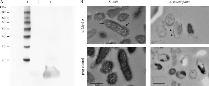
FIG 8.
Analysis of LPS and lipid A contents of A. muciniphila. (A) Western blotting of whole-cell lysates of A. muciniphila (lane 2) and E. coli (positive control) (lane 3) using an antiserum against E. coli LPS. Lane 1, molecular mass standard. (B) Electron micrographs of thin-sectioned and immunostained A. muciniphila and E. coli bacteria. The bacteria were immunostained using an antiserum against E. coli lipid A and 10-nm colloidal gold particles conjugated to protein A (pAg). Arrows indicate 10-nm gold particles. Bars, 500 nm.

