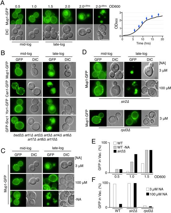
Figure 1. Niacin depletion at late-log phase induces down-regulation of cell surface proteins via transcriptional modulation by Sir2 and Rpd3.
A) Wild-type cells expressing Mup1-GFP were grown in minimal media lacking methionine (SD-Met) and imaged at different stages of growth. The final time points (+2 and +6) were 2 or 6 hrs after the cell culture had reached OD600 2.0. Right, corresponding growth curve of the culture with sampled time points indicated.
B) Localization of GFP-tagged cargo proteins in mutant cells lacking 9 known Rsp5 adaptor proteins grown to mid- and late-log phase.
C) Cells expressing Mup1-GFP were grown to mid- and late-log phase in SD-Met media with 3 µM NA, 100 µM NA, or SD-Met media without NA.
D) Mup1-GFP localization in sir2Δ and rpd3Δ null mutant cells in SD-Met media containing indicated levels of NA.
E) Quantitation of intravacuolar Mup1-GFP localization from microscopy shown in C and D. Quantified is the proportion of cells (n = >150) showing GFP fluorescence in the vacuole.
F) Percentage of Mup1-GFP expressing cells (n = >130) showing vacuolar GFP in WT, sir2Δ, and rpd3Δ cells grown in standard SD-Met media with 3 µM or 100 µM NA. Bar = 5 µm.

