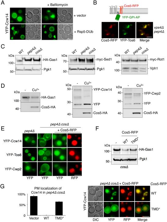
Figure 7. MVB sorting of GPI-anchored proteins is Cos-dependent.
A) Localization of YFP-Ccw14 in wild-type cells carrying a vector control or expressing Rsp5-DUb. Cells were also imaged 3 hrs after the addition of 1 µM Bafilomycin.
B) Co-localization of Cos5-RFP and YFP-Tos6 in vps4Δ pep4Δ cells.
C) Cells (WT, pep4Δ, cosΔ) expressing HA-Gas1, myc-Sed1 or myc-Rot1 were grown to log phase, prior to immunoblot analysis with α-HA or α myc antibodies and α-PGK or α-Vph1 blots for loading control.
D) The levels of GPI-APs (HA-Gas1, YFP-Ccw14 or YFP-Cwp2) in cells co-expressing Cos5-HA were measured by immunoblotting. Cultures were grown in the presence of copper chelator (−) or 50 µM copper chloride (Cu2+). In the case of YFP-tagged proteins, the vacuolar-processed form of the protein was also detectable (YFP).
E) Sorting of YFP tagged GPI-APs was assessed by fluorescence microscopy in pep4Δ and pep4Δ cosΔ cells. Defective sorting observed in cosΔ cells was restored upon expression of Cos5-RFP from a CUP1-Cos5-RFP plasmid.
F) Levels of HA-Gas1 are shown from wild-type cells co-expressing vector (−), Cos5-RFP (WT) and Cos5TMD*-RFP (TMD*). PGK immunoblot was used as a loading control.
G) Mutant cosΔ cells co-expressing YFP-Ccw14 and either WT or TMD* versions of Cos5-RFP. Left, the number of cells with YFP signal detected at the cell surface was quantified for each condition (average + SD). Bar = 5 µm.

