Fig. 1.

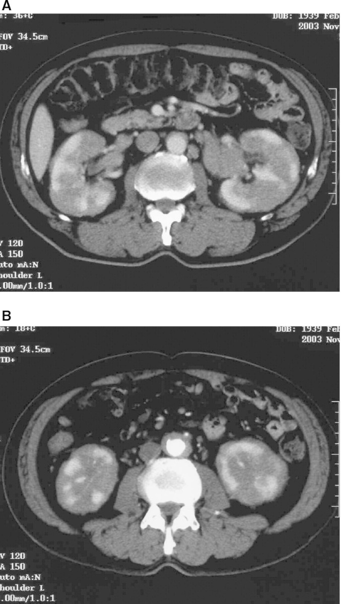
CT imaging at referral. (A) Both kidneys were markedly enlarged and showed irregular contrast staining. The pancreas was normal morphologically. (B) A paraaortic low-density area was considered to be retroperitoneal fibrosis.

CT imaging at referral. (A) Both kidneys were markedly enlarged and showed irregular contrast staining. The pancreas was normal morphologically. (B) A paraaortic low-density area was considered to be retroperitoneal fibrosis.