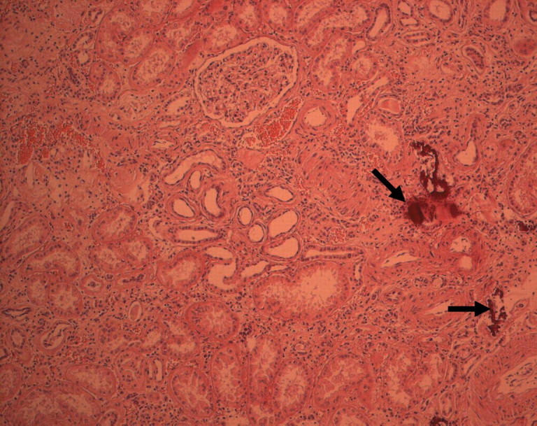
Fig. 5.

Histological examination of the left kidney. The kidney tissue surrounding the renal clear cell carcinoma shows important interstitial fibrosis and tubular atrophy. There is an interstitial lymphocytic infiltrate. Several interstitial calcifications are present (arrows).
