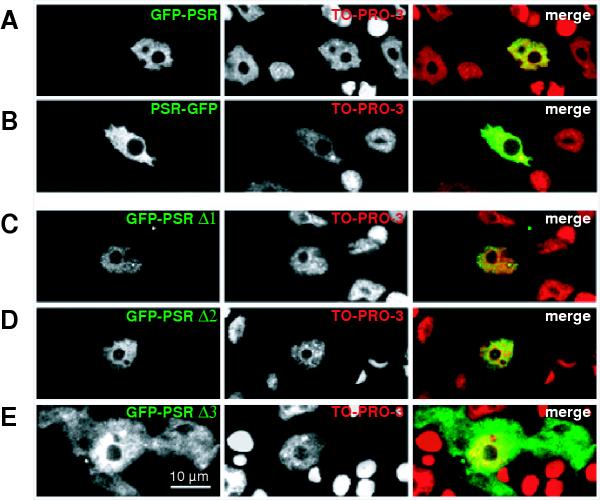
Figure 5.
PSR localisation in fixed animals Single optical sections of GFP-PSR (A), PSR-GFP (B) and single NLS-mutants ΔNLS 1, 2 and 3 in single GFP expressing cells after fixation (C-E). Left hand panels detect GFP, middle panels DNA-staining with TO-PRO and right hand panels represent merged images. Nuclear morphology is typical for hydra epithelial cells after fixation. Note that nucleoli are not stained with TO-PRO (middle panel) and also free of GFP (left hand panel).

