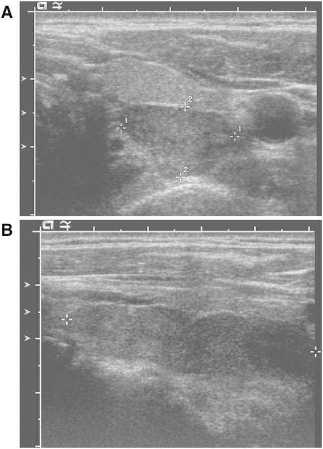
Fig. 1.
Ultrasound examination of the thyroid gland. Ultrasound of the thyroid showed a normal thyroid gland with a 3.7 × 1 × 1.3-cm left lobe and a 4.4 × 1.3 × 1.8-cm right lobe that were unremarkable in appearance. A large, elongated, hypoechoic, hypervascular mass with internal cystic change was found behind the entire left lobe of the thyroid. It measured 1.7 × 1 cm in the transverse plane (A, crosshairs 1 and 2, respectively) and ~5 cm in the longitudinal plane (B, crosshairs), suggesting either a massive single parathyroid adenoma or separate adenomas immediately adjacent to each other.

