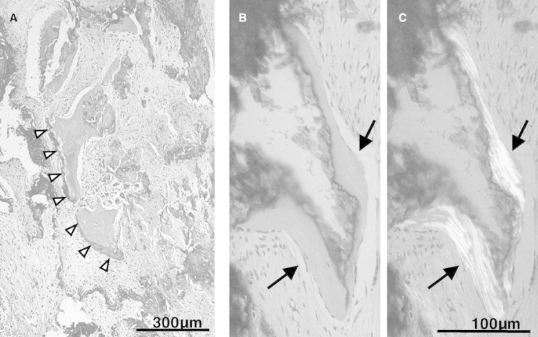
Fig. 3.

Microscopic findings of the sclerotic dura mater. (A) Calcified tissues were found in such sclerotic lesions with a rock barnacle-like appearance. Cells forming a single layer were found on the surface of this tissue (arrow heads). (B and C) On polarization microscopic observation, a clear lamellar structure was confirmed in the calcified tissue (arrows).
