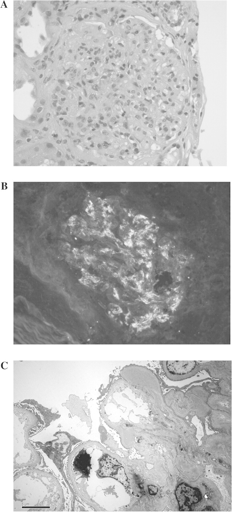
Fig. 2.

(A–C) Renal biopsy findings: (A) light microscopy with endothelial and mesangial cell proliferation with leukocyte infiltration. (B) Immunofluorescence staining for IgA in a granular pattern and mesangial deposition. (C) Electron microscopy with mesangial electron-dense deposits.
