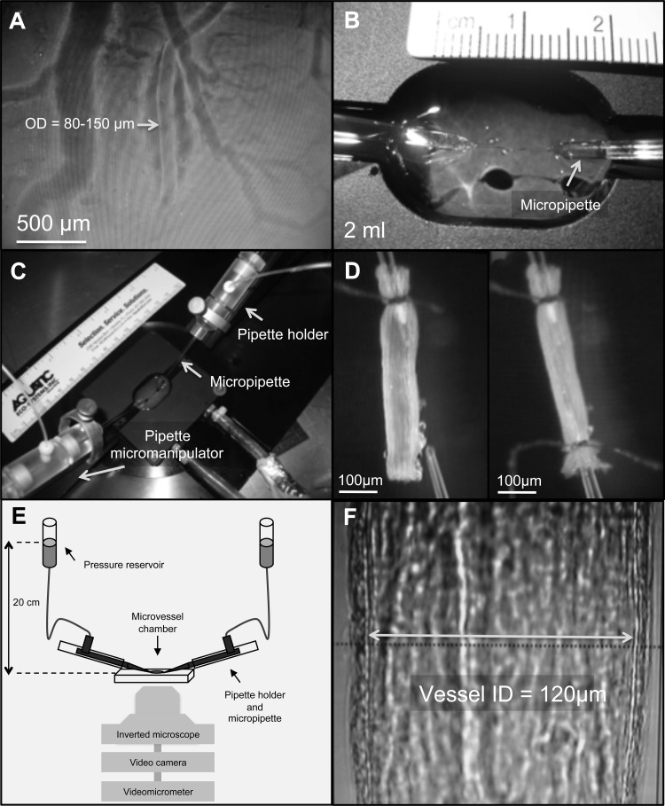
Fig. 1.
Isolation and cannulation of coronary microvessels, and methods used to study their vasoactivity. A: an unbranched arteriole [∼80–150 μm outside diameter (OD) and 1–1.5 mm long] on the surface of the ventricle. B: custom-made vessel bath containing saline solution at the experimental temperature (1°C, 5°C, or 10°C). C: vessel chamber is attached to a specially designed stage equipped with micromanipulators that house pipette holders and allowed for adjustments in pipette positioning in all three dimensions. D: arteriole was cannulated by pulling one end of the vessel onto the tip of an angled glass micropipette filled with saline (with 1% albumin) and securely tied to the pipette. After any remaining blood was flushed out, the other end of the vessel was cannulated using a second micropipette and tied in place. E and F: vessel stage was then transferred to an inverted microscope coupled with a video camera and videomicrometer system for continuous measurement of the vessel internal diameter.

