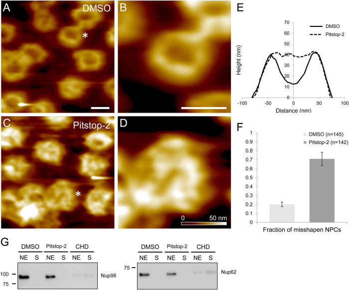
Figure 3. Pitstop-2 permeabilizes NPCs by disrupting the NPC ultrastructure without dissociating putative barrier-forming FG-nups.
AFM images of Xenopus laevis nuclear envelopes treated with DMSO (A, B). Typical appearance of the intact NPCs (A), asterisk marks an NPC magnified in panel (B). AFM images of Xenopus laevis nuclear envelopes treated with Pitstop-2 (C, D). Magnified image of an NPC marked by asterisk in panel (D). Scale bars = 100 nm. Mean section profile of 80 DMSO-treated NPCs overlaid with the mean section profile of 80 Pitstop-2-treated NPCs demonstrate an occlusion of the NPC central channel in response to Pitstop-2 treatment (E). Morphometric quantification of DMSO-treated NPCs vs. Pitstop-2-treated NPCs demonstrate a drastic increase in the fraction of misshapen NPCs in Pitstop-2-treated preparations (F). Channel depth of >10 nm was used as a criterion to differentiate between an intact or misshapen NPC. Western-blots of Xenopus leavis nuclear envelopes against putative barrier-forming FG-nups (G) show no detectable Pitstop-2-induced dissociation of either Nup62 or Nup98 (NE – nuclear envelope; S – supernatant). Imaging experiments were performed using oocytes from two different animals in duplicates. Western blot samples were prepared from two different animals in triplicates.

