
Figure 2. Topography of the graphene flake, exfoliated on top of NS-SiO2.
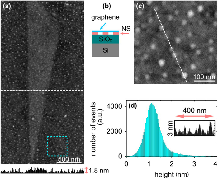
(a) Atomic force microscopy (AFM) image and a corresponding height profile along the dotted white line. (b) Side-view schematic of the graphene on top of NS-SiO2. (c) Zoomed-in AFM topography of the area inside the dotted cyan square in image (a). (d) Height histogram of the data in the image (c); inset shows a height profile along the white dotted line in the image (c).