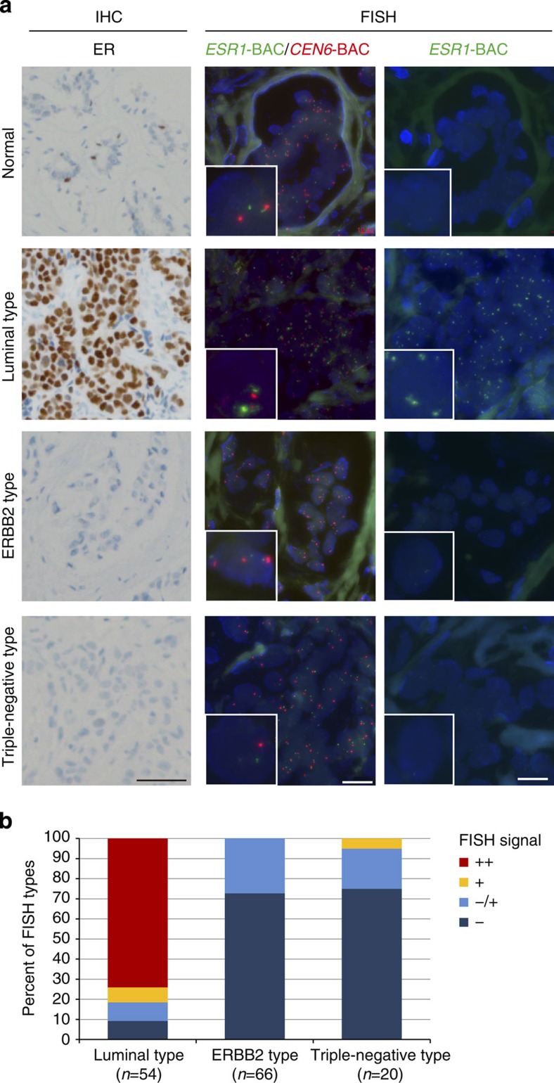
Figure 3. Eleanor expression in ER-positive breast cancer tissues.
(a) IHC and FISH analyses of breast cancer tissues. Serial sections of breast cancer tissues with the indicated types were subjected to IHC using an anti-ER antibody (IHC, left) and FISH using BAC probes for ESR1 and CEN6 (middle and right, respectively). Large Eleanor-containing foci were detected in the luminal type (ER positive). The DNA was processed with or without heat denaturation (middle and right, respectively). Scale bars, 50 μm (left) and 20 μm (middle and right). (b) Summary of FISH analyses of breast cancer patients. Strong FISH signals (++) were exclusively present in a subset of ER-expressing breast cancers (luminal type). Detailed data are provided in Supplementary Table 4.

