Figure 4.
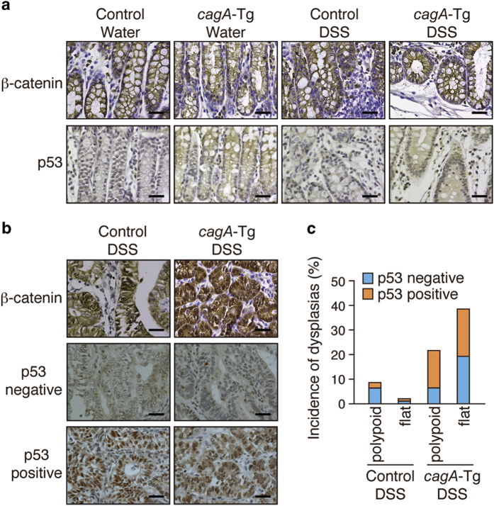
Immunostaining of β-catenin and p53 in DSS-induced dysplasias. (a) Immunostaining of β-catenin and p53 in the colonic mucosa of cagA-Tg or control mice with or without DSS treatment. Scale bars, 40 μm. (b) Immunostaining for β-catenin and p53 in dysplastic lesions of the colon. In the p53-immunostaining samples, both nuclear p53 staining-negative and -positive cases are shown. Scale bars, 40 μm. (c) Quantitative analysis of p53-negative and p53-positive dysplasias in DSS-treated cagA-Tg (n = 47) and control (n = 91) mice.

