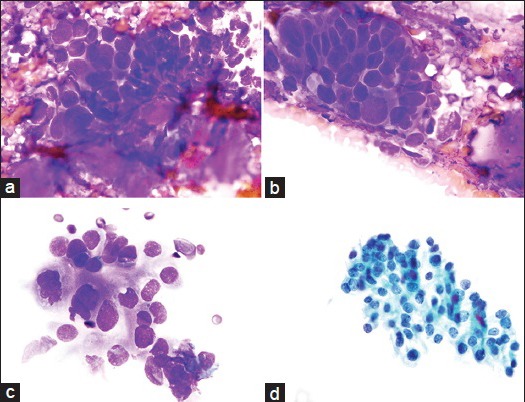
Figure 1.

(a) Group of cohesive and pleomorphic tumor cells associated with a small amount of metachromatic fibromyxoid stroma with a vague fibrillary quality. This was a focal finding (Diff-Quik [DQ] stain, ×100). (b) A cluster of cohesive and three-dimensional pleomorphic tumor cells showing small fragments of homogeneous metachromatic fibromyxoid stroma in the background. The nuclei are enlarged, hyperchromatic and angulated. Nucleoli are not evident (DQ stain, ×100). (c) Loosely-cohesive cluster of tumor cells with large, somewhat irregular nuclei with occasional nucleoli (top of the group). Cytoplasm is scant-moderate and ill-defined with a finely granular texture (DQ stain, ×100). (d) Adenocarcinoma cells on a ThinPrep (TP) display coarse irregularly clumped chromatin, occasional nucleoli and dense to granular cytoplasm (TP, Pap stain, ×100)
