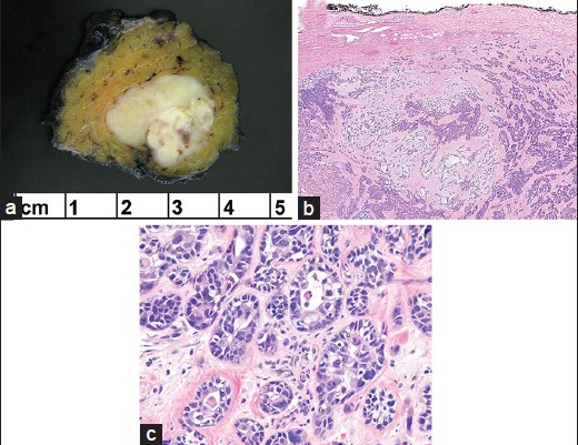
Figure 2.

(a) The resection specimen showed a well-demarcated tumor with a yellow-white cut-surface and minute areas of hemorrhage. (b) Low magnification of carcinoma ex pleomorphic adenoma (PA) admixed with PA. Note the uninvolved intact capsule of PA (H and E, ×4). (c) High magnification reveals highly atypical ductal structures and single cells with cellular anaplasia, exhibiting pleomorphic nuclei and eosinophilic cytoplasm (H and E, ×40)
