
Figure 2.
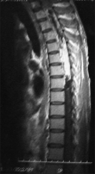
MRI sagittal section T1 weighted image with gadolinium contrast shows homogeneous intense enhancement of the solid component and irregular peripheral enhancement of the cystic part
Official websites use .gov
A
.gov website belongs to an official
government organization in the United States.
Secure .gov websites use HTTPS
A lock (
) or https:// means you've safely
connected to the .gov website. Share sensitive
information only on official, secure websites.
MRI sagittal section T1 weighted image with gadolinium contrast shows homogeneous intense enhancement of the solid component and irregular peripheral enhancement of the cystic part