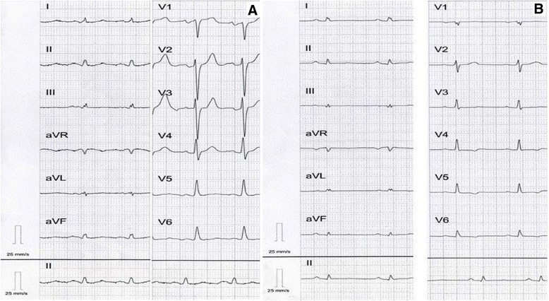
Figure 3.

Standard 12-lead electrocardiogram in the proband (A) and his daughter (B). Regular sinus rhythm in both subjects, low QRS voltage in limb leads (A), and in all leads (B). In addition, in the proband (A) ST-T changes in inferolateral leads as well as left atrial enlargement. Diffuse ST-T changes were identified in the probands’s daughter (B).
