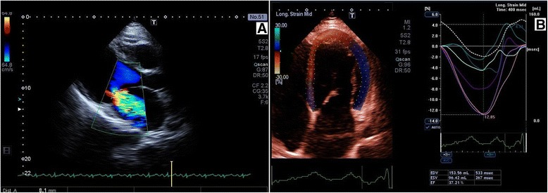
Figure 4.

Two-dimensional echocardiographic study of proband III-1. A: Parasternal long axis view in systole with color flow Doppler. Severe mitral valve regurgitation (vena contracta of 8 mm) due to restriction of the mitral valve leaflets. B: Apical four-chamber view, speckle tracking method. Enlarged left ventricle, left ventricular end-diastolic volume (LVEDV 154 ml) with low ejection fraction (LVEF 37%). Enlarged left atrium chamber.
