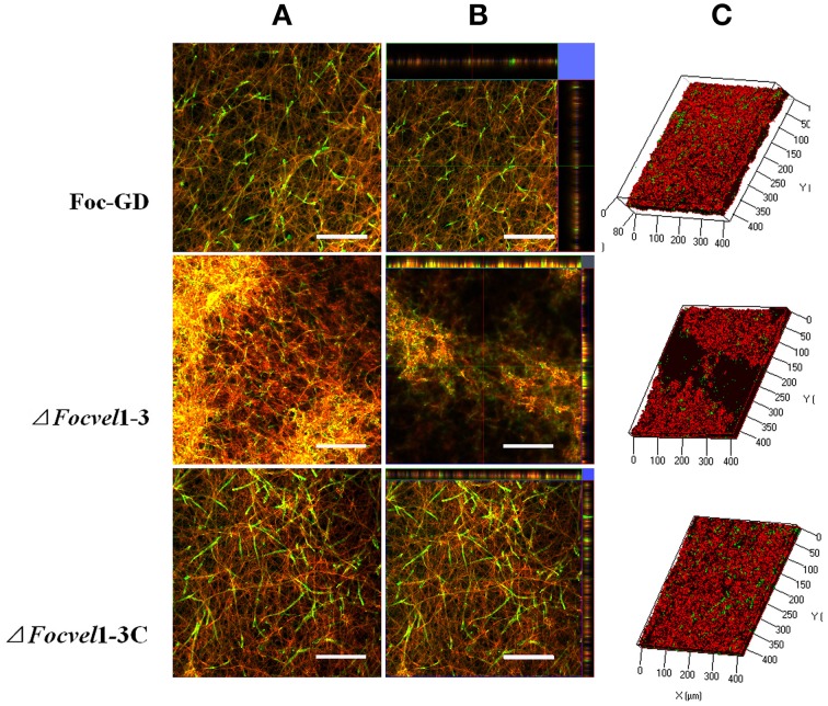
Figure 7.
CLSM images of biofilm formation by the wild-type strain (Foc-GD), FocVel1 deletion mutant (ΔFocVel1-3), and complemented strain (ΔFocVel1-3C) at 48 h. Cell wall-like polysaccharides and heat-killed biofilm cells were marked with green and red fluorescence by ConA and PI, respectively. (A) Double-stained mature biofilms. (B) Lateral views of the three-dimensional images. (C) Three-dimensional reconstruction of biofilms after dual staining. Scale bar: 100 μm.

