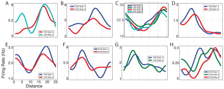
Figure 3. Examples of path equivalent cells.

(A) A cell from patient 1’s cingulate cortex during clockwise movement. (B) A cell from patient 2’s entorhinal cortex during counterclockwise movement. (C) A cell from patient 2s entorhinal cortex during clockwise movement. (D) A cell from patient 5’s entorhinal cortex during clockwise movement. (E) A cell from patient 10’s parahippocampal gyrus during clockwise movement. (F) A cell from patient 12’s entorhinal cortex during counterclockwise movement. (G) A cell from patient 13’s hippocampus during clockwise movement. (H) A cell from patient 13’s hippocampus during counterclockwise movement. See figure S3 for additional examples.
