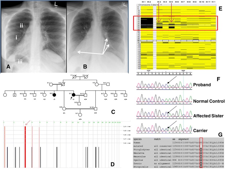
Figure 1.

Clinical and molecular genetic findings. Panel A) AP Chest X-ray of proband indicating (i) dextrocardia with (ii) right-sided aortic arch. The gastric air bubble is also in the right side (iii) that is most likely representing situs inversus. Panel B) AP Chest X-ray of proband where arrows are indicating bronchial wall thickening with mild bronchiectasis in the left middle lobe and also in the right lower lobe. Panel C) Family pedigree showing the genotypes for the p.Lys1154Gln variation in DNAH1 for all family members that were sequenced. (+) indicates the wild-type allele and (−) indicates the mutant allele. Arrow indicates the proband. Panel D) Results of analysis of genotyping data with Homozygosity Mapper, showing 4 ROHs shared among the affected individuals. The ROH on chromosome 3p23-p14.2 containing DNAH1 is indicated by an arrow. Panel E) Narrowing the single valid ROH by two unaffected family members on chromosome 3 by AutoSNPa analysis. Panel F) Sequence chromatogram of exon 20 where the mutant arrow points to the site of the c.3460 A > C transversion and Panel G) Multiple protein sequence alignment of DNAH1 orthologs showing complete conservation of the p.Lys1154 residue compared to the altered residue (indicated by red box).
