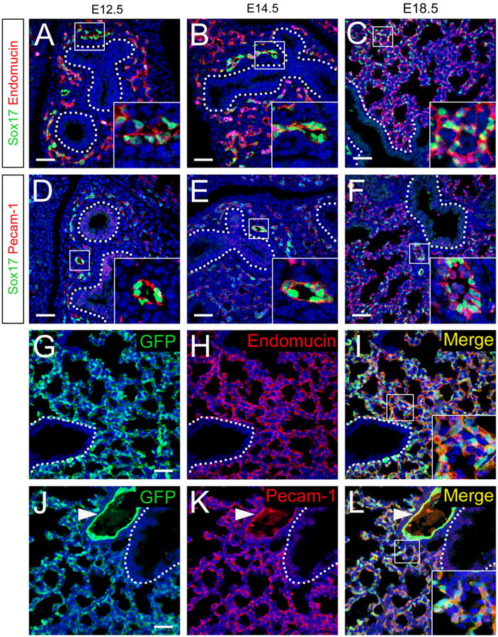
Fig. 1.

Sox17 is expressed in endothelial cells in the developing lung. (A–F) Immunofluorescence double labeling for Sox17 (green) and endomucin (red, A–C) or Pecam-1 (red, D–F) was performed on sections from E12.5 (A and D), E14.5 (B and E), and E18.5 (C and F) mouse lungs. Sox17 was detected in the nuclei of cells staining for endothelial cell markers in the developing mouse lung. (G–L) Immunofluorescence for GFP (G and J) and endomucin (H) or Pecam-1 (K) was performed on sections from E18.5 Sox17GFP reporter knock-in mouse lungs. Sox17-expressing cells labeled with the GFP reporter were co-stained with endothelial cell markers. Nuclei are stained with DAPI (blue). Insets show higher magnification of boxed regions and dotted lines denote the basal surface of the airway epithelium. Pulmonary artery (inset, F; arrowhead, J–L). Peripheral microvasculature (insets, C, I, and L). Red blood cell autofluorescence in panel F was masked using Imaris software. Scale bars 40 μm.
