
Fig. 3.
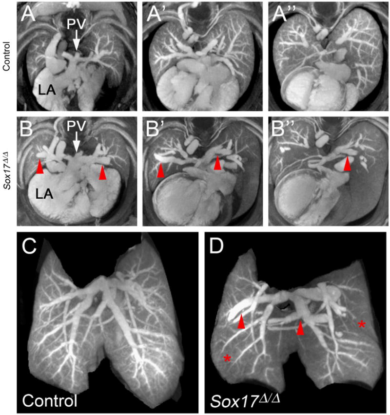
MicroCT analysis shows pulmonary vascular abnormalities in Sox17Δ/Δ embryonic lungs. (A–B) MicroCT images comprised of a merged stack of 50 2D microCT slices. Panels depict a sequential series through the developing lungs of E18.5 control (A–A″) and Sox17Δ/Δ (B–B″) mice. Pulmonary veins were dilated (red arrowheads) and vascular branching was aberrant in the lungs of Sox17Δ/Δ embryos. (C–D) Static 3D images of the developing pulmonary vascular network in E18.5 control (C) and Sox17Δ/Δ (D) lungs showing dilated pulmonary blood vessels (red arrowheads) and reduced staining of the peripheral microvasculature (red asterisks). PV, pulmonary vein; LA, left atrium.