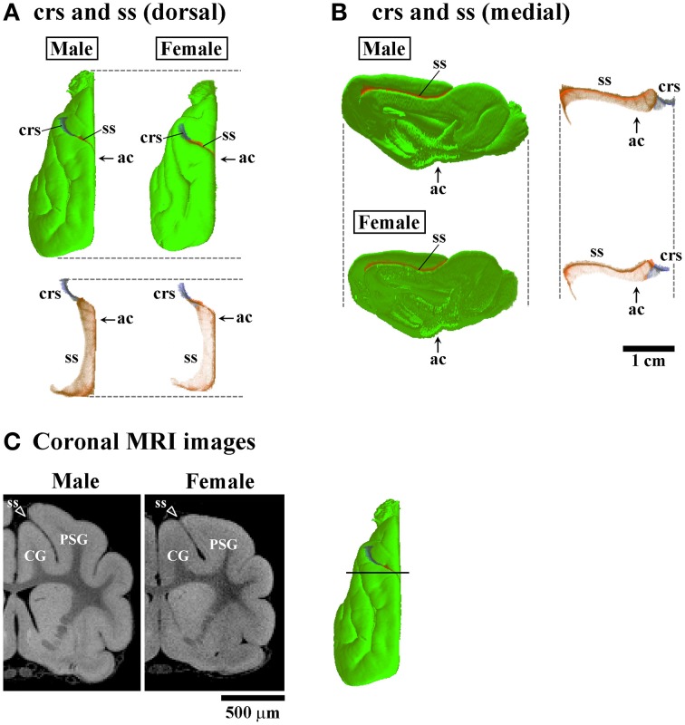
Figure 5.
Three-dimensional rendered images of crucinate and splenic sulci. (A) Dorsal views of 3D-rendered images of the crucinate sulcus (crs) and splenic sulcus (ss) with or without the cerebral surface. Each image is registered at the anterior commissure (ac). (B) Medial views of 3D-rendered images of the crs and ss with or without the cerebral surface. Each image is registered at the ac. (C) Coronal T1-weighted (short TR/TE) MRI of the ferret cerebrum corresponding to the rostral peak of ss infolding in the frontal region (Figure 3B). Bar in 3D-rendered images of the cerebrum indicates the positions of coronal MRI images acquired. The ss demarcates the cingulate gyrus (CG) and posterior sigmoid gyrus (PSG), male-prominent ss infolding result in a greater convolution of the PSG in males than in females.

