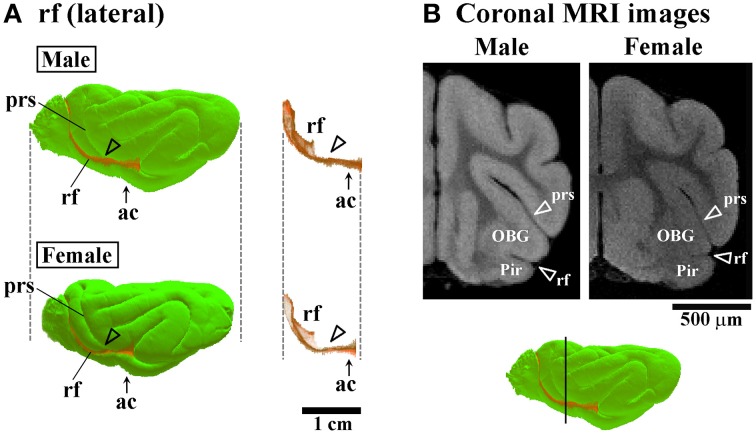
Figure 6.
Three-dimensional rendered images of rhinal fissure. (A) Lateral views of 3D-rendered images of the rhinal fissure (rf) with or without the cerebral surface. Arrowheads indicate the rf region, corresponding to a second peak of the lf infolding (see open arrowheads in Figure 4C). Each image is registered at the anterior commissure (ac). (B) Coronal T1-weighted (short TR/TE) MRI of the ferret cerebrum corresponding to the rf region, which is more infolded in males than in females in the frontal region. Bar in 3D-rendered images of the cerebrum indicates the positions of coronal MRI acquired. The ss demarcates the cingulate gyrus (CG) and piriform cortex (Pir), male-prominent rf infolding result in a greater convolution of the Pir in males than in females.

