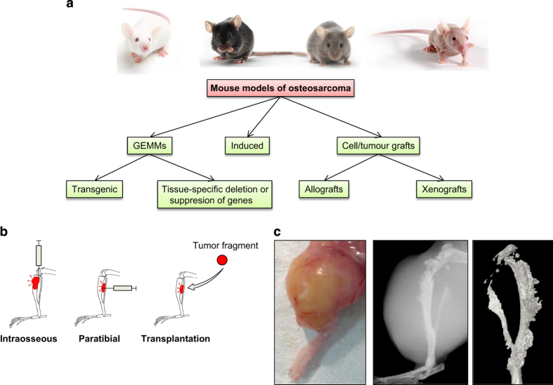
Figure 1.
Summary of models of osteosarcoma. (a) A summary of the available mouse models for OS. (b) Illustration of the different injection modes. (c) Picture taken after the skin removal of a tumor obtained 1 month after injection of KHOS cells. Radiography of the tibia carrying the tumor. Three-dimensional reconstruction of the tumoral tibia after microCT analysis.

