
Figure 1.
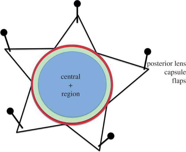
The eye lens and the different regions within the lens epithelium. The lens epithelium can be subdivided into two distinct regions, a central and a peripheral region. The latter comprises two zones called the germinative (GZ) and transitional (TZ) zones. When the anterior lens capsule is flat mounted with the epithelial cells exposed after the removal of the lens fibre mass and the dissected portions of the posterior lens capsule pinned into place, then these regions are apparent. The anterior pole is indicated (+). The central region (blue) is the largest and it is where cell proliferation occurs at a low basal rate. The cells in this region are flatter and less densely spaced. Cell proliferation is largely restricted to the peripheral region and in particular the GZ (green). Proliferating cells were first identified by observing mitotic figures and their incorporation of tritiated thymidine, but now the incorporation of a thymidine analogue such as BrdU or the nucleoside EdU is used. Alternatively, the immunodetection of Ki67, a marker of cells in S-phase, or PCNA is used. Progeny from the GZ cells become lens fibre cells by migrating centripetally towards the lens equator and passing through the TZ and MR (red), before exiting the epithelium via the MR into the body of the lens. MR cells are considered post-mitotic. Cells in the GZ, TZ and MR, comprising the peripheral region of the lens, are shielded from light, but not IR, by the iris and are out of the visual axis.