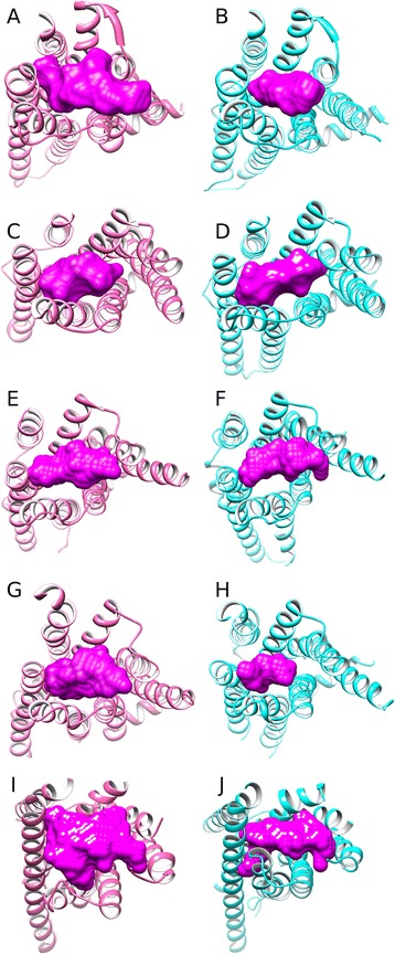
Figure 10.

A comparison of inactive states (in pink) and active states (in cyan) of Class A GPCRs (extracellular view): (A) and (B): adenosine A2A receptor, (C) and (D): rhodopsin, (E) and (F): β2-adrenergic receptor, investigate pathways of activation(G) and (H): muscarinic acetylcholine M2 receptor, (I) and (J): P2Y purinoceptor12. Binding-site volumes (in magenta) were calculated with POVME [52].
