
Figure 12.
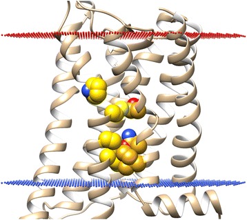
TM6-TM7 interface of Rhodopsin in its inactive state (PDB id: 1GZM) with residue positions 6.40, 6.43, 6.47, 6.50, 7.45, 7.49, 7.52 highlighted (in yellow/gold) (Ballesteros-Weinstein numbering [60]). Upon mutation these “hotspot” residues increase constitutive activity of some Class A GPCRs by destabilising their inactive states. Extracellular side of membrane is represented by red dots and the intracellular side by blue dots.