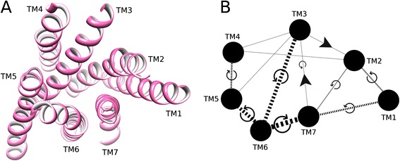
Figure 2.

Helix pairs in Class A GPCRs. (A) Extracellular view of 7TM fold of the muscarinic acetylcholine M2 receptor (PDB id: 3UON). (B) 2D representation of average conformational changes during receptor activation (from 18 inactive and 7 active Class A GPCRs). Helices are solid black circles. Helix pairs connected with black dotted lines. Line width is proportional to change in interhelical angle. Circular arrows give angle rotation direction: anticlockwise for angle decrease (helices become more parallel), clockwise for angle increase (helices become less parallel). Solid black arrows show translational movement of helices.
