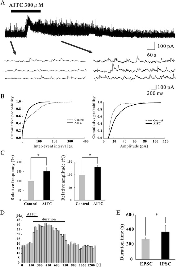
Figure 2.

Actions of AITC on inhibitory synaptic transmission in SG neurons. (A) Continuous chart recording of IPSCs before and during the action of AITC (300 μM; top). Three consecutive traces of IPSCs are shown in an expanded scale of time, before (bottom left) and under the action of AITC (bottom right). The holding potential (VH) used was −0 mV. (B) Cumulative distribution of the inter-event interval (left) and amplitude (right) of IPSCs in the control (dotted line) and during (continuous line) the action of AITC. AITC shifted the inter-event interval and amplitude to a shorter and a larger one (same neuron as in Figure 2A). (C) Summary of IPSC frequency (left) and amplitude (right) under the action of AITC (n = 9) relative to control. (D) The frequency of IPSCs following application of AITC plotted against time. Each bar indicates data calculated from IPSCs measured for 30 s (same neuron as in Figure 2A). “Duration” represents the period when frequency increases more than 20% of the control. (E) Summary of average duration of IPSCs and EPSCs evoked by AITC (n = 5). *P < 0.05.
