
Figure 3.
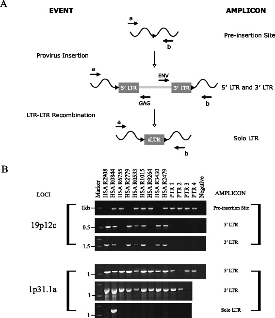
Detection of structural variation at HERV-K(HML-2) loci. (A) Formation of alleles and strategy for independent amplification of structural variations in novel human HERV-K(HML-2) proviral loci. Primer binding sites are indicated by arrows ( ). The locus specific primer combination a + b amplifies the pre-insertion site and solo LTR. Primer combination a + GAG amplifies the 5′ LTR of a full-length provirus and ENV + b the 3′ LTR. (B) Composite agarose gels of locus specific amplification for allelic variation. Numbers on the left correspond to the co-migrating DNA size marker (kb). Samples are the same as used in Figure 1D, with humans in a different order. The identity of each band was confirmed by DNA sequencing. Refer to Additional file 7 for un-cropped gel images.