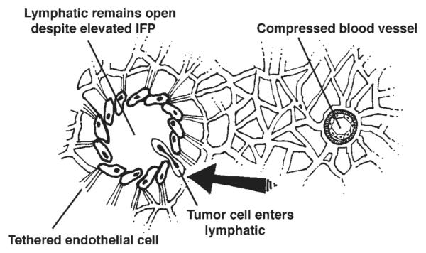
Fig. 5.

The microstructure of the lymphatic wall (on the left), with patchy basement membrane, tethering of endothelial cells to surrounding stroma, and “button-like” interendothelial openings [13] remains open to the inflow of fluid and tumor cells despite increased IFP. In contrast, the collapsed blood vessel (right) probably is more difficult for a tumor cell to negotiate.
