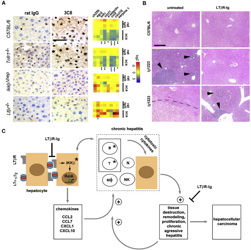
Figure 8. Effects of acute 3C8 and long-term LTβR-Ig treatment and a model of chronic inflammation-induced hepatocarcinogenesis in tg1223 mice.
(A) Immunohistochemical analysis of nuclear p65 translocation and real-time PCR for mRNA expression of selected NF-κB target genes in livers of C57BL/6 and various knock-out mice treated with 3C8. Data are presented on a log 2 scale (blue: upregulated; red: downregulated). Rows indicate individual mice; columns represent particular genes. Each data point reflects the median expression value of a particular gene resulting from 3–4 technical replicates, normalized to the mean expression value of the respective gene in C57BL/6 livers. (scale bar: 50μm). Expression data are depicted according to treatment group: rat IgG (control) or 3C8 (LTβR agonist). Statistical significance was evaluated by t-test: * = p≤0.05; ** = p<0.001; *** = p<0.0001.
(B) Histological analysis (H&E) of livers from untreated (left column) and LTβR-Ig treated (right column) C57BL/6 or tg1223 mice (12 months of age). Representative sections show no hepatitis or HCC in untreated or LTβR-Ig-treated C57BL/6 livers (upper row). Untreated tg1223 livers display hepatitis in 34/34 (middle panel, left column) and HCC in 6/34 cases (lower panel, left column). LTβR-Ig treatment reduces the incidence of hepatitis (middle and lower panel, right column) and prevents HCC formation in LTβR-Ig treated tg1223 mice. Arrowheads indicate inflammatory foci. Tumor border is indicted by a dashed line (scale bar: 200μm).
(C) Scheme of chronic inflammation-induced liver carcinogenesis in tg1223 mice: Transgenic hepatocytes (brown) express LTα, β and induce chemokine production (e.g. CCL2, CCL7, CXCL1, CXCL10) in the presence of IKKβ and intrahepatic lymphocytes. Chemoattraction and activation of myeloid cells and lymphocytes expressing particular chemokine receptors (e.g. CXCR3, CXCR2, CCR2, CCR1) causes hepatitis: CXCL10 attracts CXCR3+ T- and NK-cells, CXCL1 CXCR2+ T-, B-cells and neutrophils, CCL2 CCR2+ macrophages and CCL7 attracts CCR1+ monocytes. Activated, infiltrating immune cells secrete cytotoxic cytokines (e.g. IL6; IL1β, TNFα, IFNγ, LTαβ) that cause tissue destruction, hepatocyte proliferation, cell death and tissue remodeling. In such an environment, hepatocytes are susceptible to chromosomal aberrations leading to HCC. Tissue destruction and remodeling supports the infiltration of activated inflammatory cells (e.g. myeloid cells) leading to a feed-forward loop towards chronic aggressive hepatitis. Asterisks indicate that genetic depletion of those components (IKKβ; T- and B-cells) blocks chronic hepatitis development and HCC. Blocking LTβR signaling with LTβR-Ig in 9 month-old tg1223 mice reduces chronic hepatitis incidence and prevents HCC. +: indicates the fortification of a described process. ┤: indicates the suppression of a described process. The transcription factor RelA is schematically depicted as a green circle, inducing transcription of NF-κB target genes (e.g. chemokines) (arrow). B, T: B- and T-cells. MØ: macrophages. N: neutrophils. NK: NK-cells.

