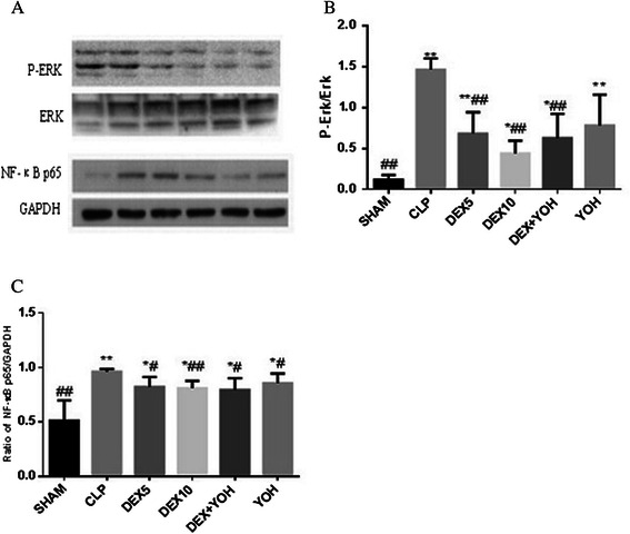
Figure 3.

Western blot analysis of P-ERK and NF-κB p65. Rats were treated with dexmedetomidine (5 μg/kg or 10 μg/kg), or yohimbine(1.0 mg/kg) for six hours after CLP or operation. P-ERK and NF-κB p65 activation in lung tissues were analyzed by Western blot (A); The correspondingly gray intensity analysis of the western blot were shown in (B) and (C). Data were presented as the mean ± standard deviation. *P < 0.05 and **P < 0.01 vs. the sham group;#P < 0.05 and ##P < 0.01vs. CLP group,n = 8.
