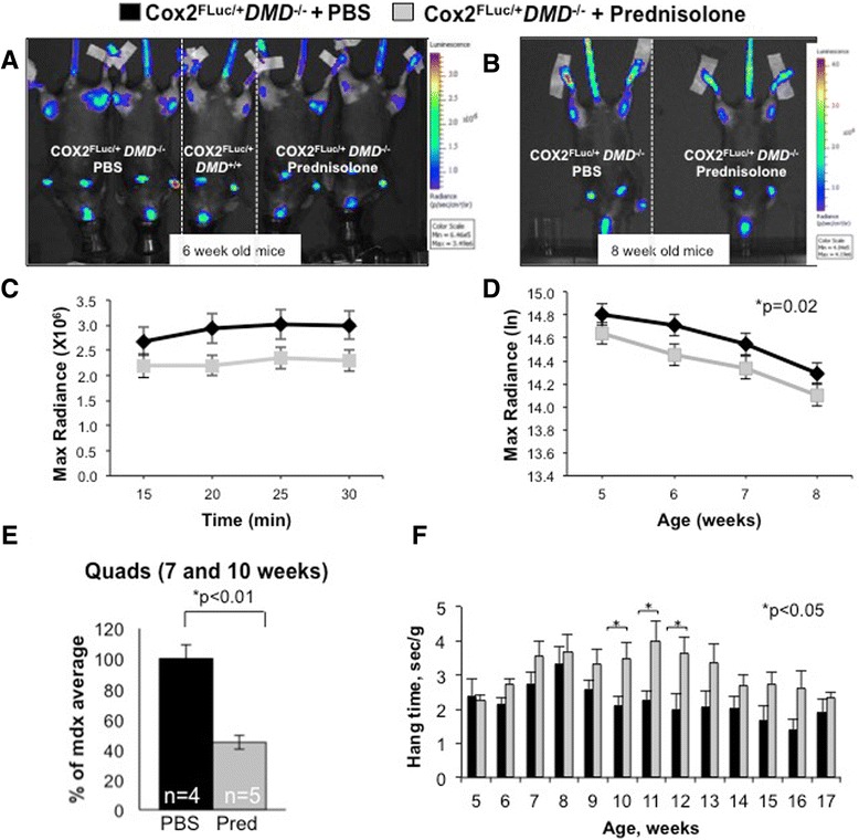
Figure 4.

Bioluminescent signal is reduced in prednisolone-treated Cox2 FLuc/+ DMD −/− mice. Mice were treated with 0.75 mg/kg of prednisolone or with PBS for 5 days a week starting at 2 weeks of age. Imaging was initiated at 5 weeks of age. (A) Representative bioluminescent images taken 25 min after D-luciferin injection into prednisolone-treated Cox2 FLuc/+DMD−/− mice at 6 weeks of age (two mice, far right), PBS-treated Cox2 FLuc /+DMD−/− mice (two mice, far left), a control Cox2 FLuc/+ DMD +/+ mouse (one mouse, middle), and (B) representative images from mice imaged at 8 weeks of age (PBS treated on left, prednisolone treated on right). All mice were females. (C) Representative example of one imaging time point (6 weeks of age) showing the stability of the signal over the 30 min imaging period. (Prednisolone, N = 37; PBS N = 39). (D) Optical imaging data from prednisolone (N = 35, 37, 38, 34) and PBS (N = 33, 39, 37, 37) treated Cox2 FLuc/+ DMD −/− mice from 5 to 8 weeks of age. Bars represent standard errors. Data are expressed as max radiance and graphed as log base e, (E) luciferase assays of quadriceps muscles harvested from 7 to 10-week-old prednisolone and PBS-treated Cox2 FLuc/+ DMD −/− mice. (F) Wire strength test of prednisolone-treated (N = 29, 24, 21, 16, 17, 16, 12 15, 11, 9, 13, 7, and 7) and age-matched control (N = 25, 29, 22, 17, 17, 13, 16, 16, 14, 13, 16, 5, and 9) Cox2 FLuc/+ DMD −/− mice for ages 5, 6, 7, 8, 9, 10, 11, 12, 13, 14, 15, 16, and 17 weeks. *P < 0.05; PBS, phosphate-buffered saline.
