Figure 2.

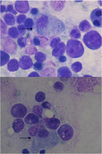
Bone marrow cytology. May-Grunwalds/Giemsa-stained bone marrow cytology. The pictures show increased bone marrow cellularity with an hemophagocytosed erythroblast (black arrow).

Bone marrow cytology. May-Grunwalds/Giemsa-stained bone marrow cytology. The pictures show increased bone marrow cellularity with an hemophagocytosed erythroblast (black arrow).