
Figure 2.
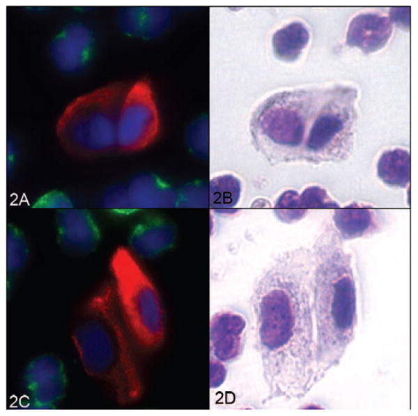
Two circulating tumor cell (CTC) clusters displayed in fluorescence and in the corresponding Wright-Giemsa stain. Left, Fluorescent CTCs (A and C). Right, The same cells after Wright-Giemsa staining (B and D) (red, cytokeratin; green, CD45; blue, 4′,6-diamidino-2-phenylindole; original magnifications ×60).