
Figure 1.
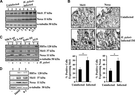
H. pylori–induced Noxa and Mcl1 expression in GECs. A) A representative Western blot (n = 3) of whole-cell lysates prepared from uninfected and infected (3, 5, 8, and 12 h with an MOI of 200) AGS cells showed time-dependent induced expression of Noxa and Mcl1. α-Tubulin was used as a loading control. B) Immunohistochemical staining of biopsy samples from uninfected and H. pylori-infected patients showing expression of Noxa as well as Mcl1 in the epithelium as well as lamina propria (open arrows indicate epithelial cell lining in the gastric mucosa). Quantification is shown of Mcl1+ and Noxa+ cells in the infected and uninfected gastric mucosa (n = 5). Graph shows the mean ± sem. *P < 0.05, Student’s t test. Original magnification, ×50. Scale bars, 100 μm. C) Expression of Hif1α, Mcl1, and Noxa was analyzed by immunoblotting cell lysates prepared from uninfected or H. pylori–infected (an MOI of 200; 5 h) MKN-45, NCI-N87, KATO III, and AGS cells. α-Tubulin was kept as a loading control. D) Western blot analysis of epithelial cells isolated from 3 sets of uninfected human gastric biopsy samples and separately infected with either an MOI of 200 of cag PAI(−) 8-1 strain or (+) 26695 strain for 5 h. IM, intestinal metaplasia.