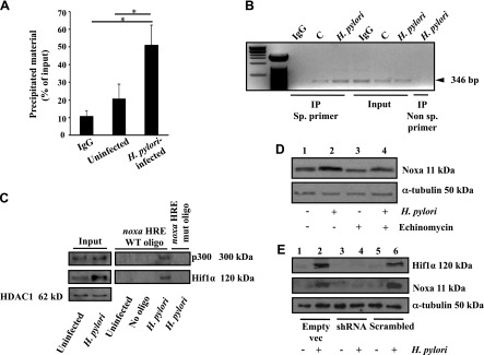
Figure 2.

Hif1α and its coactivator p300 interacted with the promoter region of noxa and enhanced Noxa expression after H. pylori infection. A) Enrichment of the noxa promoter HRE by ChIP with Hif1α antibody relative to input DNA measured by real-time PCR (mean ± sem, n = 3). *P < 0.05. B) ChIP analysis of Hif1α immunocomplex for the noxa promoter HRE. Specificity of Hif1α binding to the noxa HRE was confirmed by comparing the 5′ far upstream region (nonspecific [sp.] primer) with the HRE-flanking region of the noxa promoter. C) Western blotting (n = 3) showed binding of Hif1α and its transcriptional coactivator p300 to the noxa promoter HRE (coated on streptavidin beads) after infection with an MOI of 200 of H. pylori for 5 h. D) Hif1α-induced Noxa expression was validated by pretreating AGS cells with 150 nM Echinomycin prior to infection with an MOI of 200 of H. pylori for 5 h. α-Tubulin was the loading control. E) Western blotting (n = 3) showed expression of Noxa and Hif1α in the negative control shRNA, scrambled negative control shRNA, and Hif1α-shRNA stably expressing AGS cells. IP, immunoprecipitation; vec, vector.
