
Figure 4.
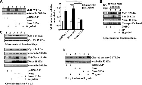
Mcl1 degradation, cytochrome c release, and cleaved caspase-3 formation were enhanced in S13A Noxa-expressing H. pylori–infected GECs. A) AGS cells were transfected with either empty vector, Noxa WT or Noxa S13A construct followed by infection for 5 h with an MOI of 200 of H. pylori, and Mcl1 status was analyzed by immunoblotting of whole-cell lysates. Data were analyzed by 2-way ANOVA with Tukey’s post hoc test (n = 4). Error bars, sem. ****P < 0.0001. B) Immunoprecipitation experiment assessing Mcl1-Noxa interaction and Mcl1-Bax interaction in the mitochondrial fraction of infected AGS cells. Mitochondrial extracts were immunoprecipitated with Mcl1 antibody after various treatments as indicated. A nonspecific band was used to estimate protein loading. C) Western blot showing phosphorylation status of Noxa and cytochrome (Cyt) c in the cytosol in 9 h H. pylori–infected AGS cells expressing either empty vector or Noxa WT or Noxa S13A. Corresponding mitochondrial fraction assessed cytochrome c retention status 9 h p.i. Cox IV was used as a loading control for mitochondrial fraction. D) Immunoblot of whole-cell lysates showing the status of cleaved caspase-3 in Noxa WT and S13A Noxa-transfected AGS cells infected with an MOI of 200 of H. pylori for 10 h. α-Tubulin was used as a loading control.