Figure 5.
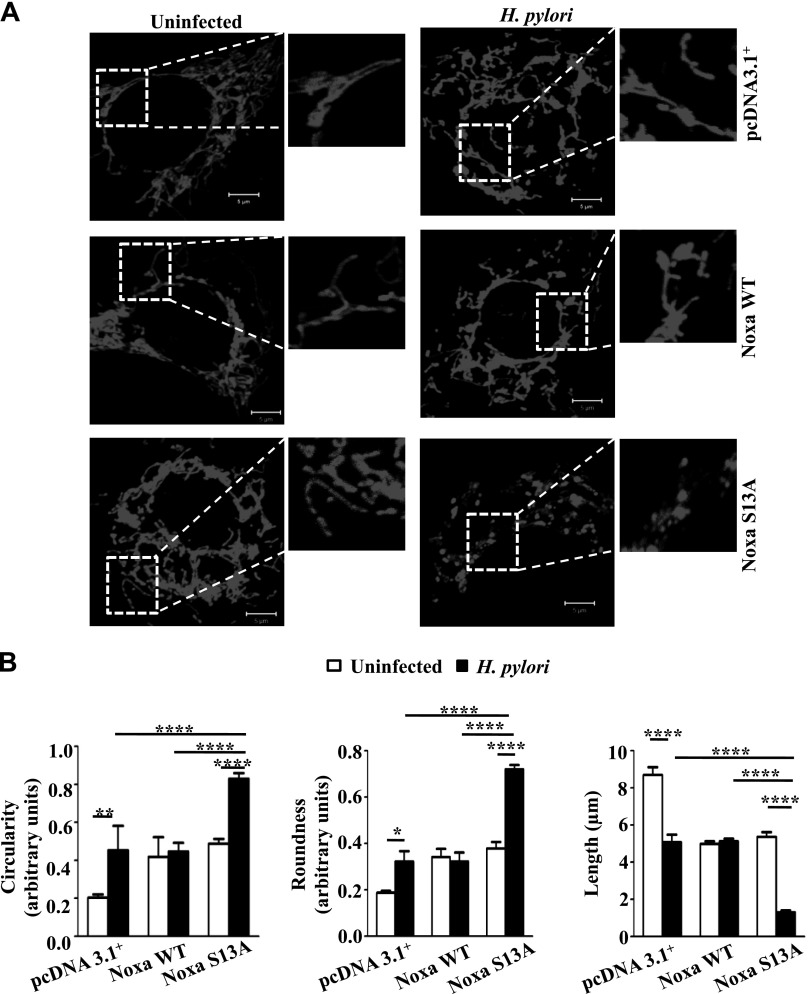
Non–P-Noxa increases H. pylori–induced mitochondrial fragmentation. A) Mitochondrial morphology was examined by confocal microscopy after transfecting empty vector (pcDNA3.1+), Noxa WT, and Noxa S13A constructs in pDsRed2 stably expressing AGS cells followed by infection with H. pylori for 8 h, with an MOI of 200. Scale bars, 5 µm. B) Mitochondrial length, roundness, and circularity information was collected from 4 cells (from 4 independent experiments), and 5 mitochondria were measured from each cell for statistical analysis. Circular mitochondrial appearance indicative of mitochondrial stress was more apparent in infected Noxa S13A-transfected cells when compared with the pcDNA3.1+or Noxa WT construct-expressing infected cells. Data were analyzed by 2-way ANOVA with Tukey’s post hoc test. Error bars, sem. *P < 0.05; **P < 0.01; ****P < 0.0001.

