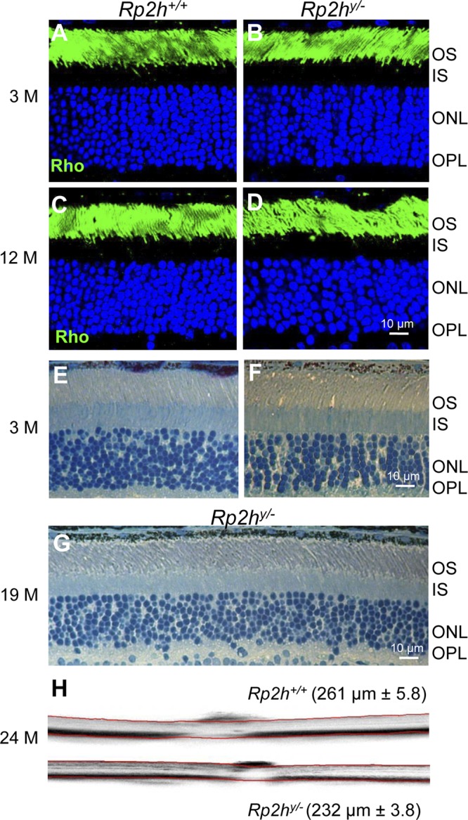
Figure 3.

Slow rate of Rp2h−/− retina degeneration. A–D) Frozen sections of WT and mutant retina were probed with anti-rhodopsin antibodies (Rho, green) at 3 mo (3M) (A, B) and 12 mo of age (C, D). E–G) Plastic sections of WT and mutant retina at 3 mo (E, F) and of a 19-mo-old Rp2hy/− breeder (G). H) OCT of 2-yr-old WT (top) and mutant (bottom) retina. OS, outer segment; IS, inner segment; ONL, outer nuclear layer; OPL, outer plexiform layer. Scale bar, 10 μm.
