
Fig. 7.
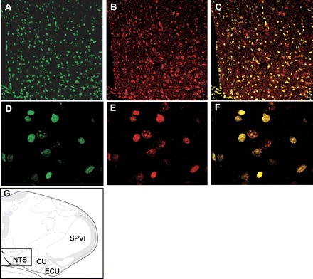
GBR1 localization and expression in neurons of the NTS. A–C: fluorescence confocal images (×10 magnification) demonstrating neurons within the NTS stained with a specific anti-GBR1 antibody (green, A) and a neuron-specific anti-NeuN antibody (red, B). C: overlap of A and B, indicating that GBR is located on neurons. D–F: fluorescence confocal images taken under high magnification (×40) from a section of the NTS brain sections shown in A–C, respectively. G: location of the stained NTS brain sections shown in A–F, based on the rat brain atlas of Swanson (37a). SPVI, spinal tract of the trigeminal interpolar part; ECU, external cuneate nucleus; CU, cuneate nucleus.