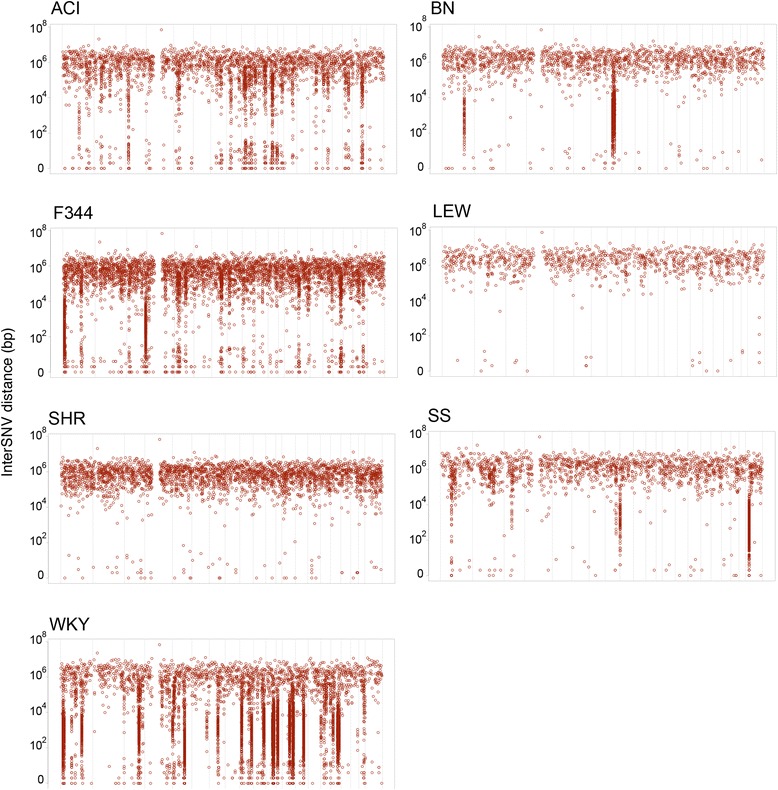
Figure 4.

Genomic distribution of substrain variants per strain. For each strain the distance between two consecutive SNVs (y-axis) is plotted along the genomic position (x-axis). The windows on the x-axis represent the different chromosomes. Loci with a high density of substrain SNVs can be observed as clusters that drop down from the average genome-wide pattern.
