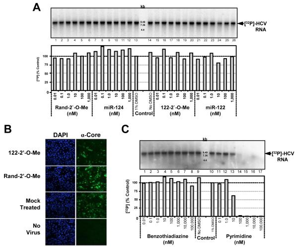
Figure 1. Effect of miR-122 on the elongation phase of HCV RNA synthesis by cell-free replicases.
(A) Various concentrations of miR-122 or other oligonucleotides (Table 1) were added to the P16 fraction from subgenomic replicon cells in the cell-free replicase reaction. Control reactions were carried out either in the absence (lane 14) or presence (lane 13) of 1% DMSO. Radiolabeled RNA was extracted, and resolved on a 1% agarose gel containing glyoxal, followed by autoradiography (top panel) or PhosphoImager quantitation (lower panel). (B) Huh-7.5 cells were transfected with 50 nM oligonucleotide, and 24 hrs later infected with HJ3-5 virus, a chimeric HCV, at an m.o.i. of 1. Cells were immunostained for core antigen 72 hrs after infection. Nuclei were labeled with DAPI. (C) Increasing concentrations of an allosteric benzothiadiazine or active-site pyrimidine inhibitor were incubated with the P16 fraction.

