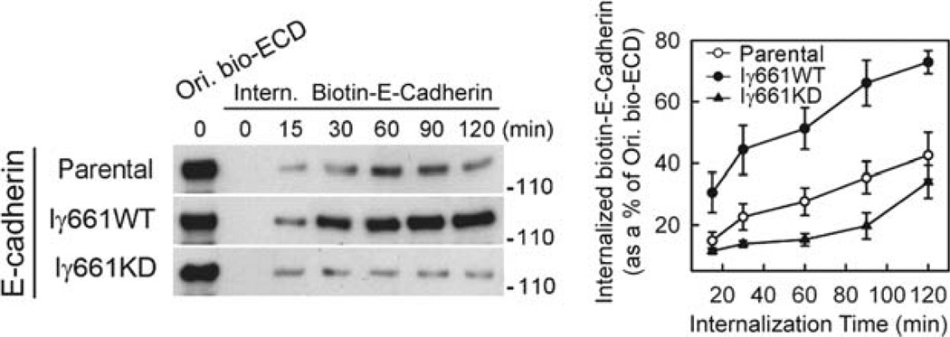
Fig. 1.
Overexpression of wild-type PIPKIγ661 accelerates E-cadherin internalization, while expression of kinase dead (KD) PIPKIγ661 impedes it (11). MDCK cells expressing PIPKIγ661 wild-type and kinase dead were biotinylated (Ori. Bio-ECD) with sulfo-NHS-SS-biotin followed by EGTA treatment to induce endocytosis. The cells were washed with glutathione to remove the disulfide linked biotin from cell surface accessible E-cadherin at intervals to monitor the rate of internalization. The cells were processed to determine total levels of biotinylated E-cadherin by western blotting. The results of the blotting were quantified (right) by densitometry using NIH ImageJ. The expression of active PIPKIγ661 clearly enhances the rapid endocytosis of E-cadherin while the kinase dead PIPKIγ661 does not.

