Figure 1.

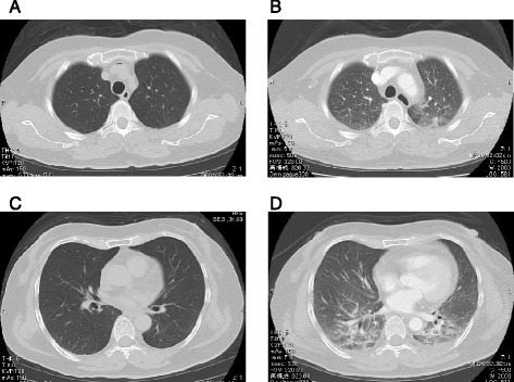
Computed tomography scans. (A, C) The plain computed tomography scan on admission. (B, D) The contrast computed tomography scan at hemodynamic deterioration. A and B were the same level of the body. C and D were the same level of the body.

Computed tomography scans. (A, C) The plain computed tomography scan on admission. (B, D) The contrast computed tomography scan at hemodynamic deterioration. A and B were the same level of the body. C and D were the same level of the body.