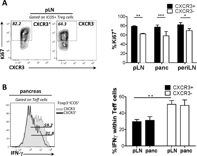
Fig 2. CXCR3 expression delineates a functionally fit subpopulation of ICOS+ Treg cells.
(A) Cell suspensions of pancreatic draining LN from 4-week-old mice were isolated and percent cycle cells, determined by Ki-67 expression, were compared between the CXCR3+ and CXCR3- subsets of ICOS+ Treg cells. (B) NOD.TCRα-/- mice received Teff (7.5X10⁵) cells and either CXCR3+ or CXCR3- ICOS+ Treg cells (7.5X10⁴) cells isolated from pooled LN and spleen of BDC2.5 mice. After 14 days suppression was assessed via percent IFN-γ+ cells among Teff cells in the pancreas and draining LN. (n = 4).

